GE Medical Engineers Apply VR Technology to Preoperative Preparation
Imagine that you are a doctor. When you enter a virtual scene, this scene turns out to be the patient's brain. At the same time, you can zoom in according to your needs. You need to know the area in detail. How amazing it will be for a doctor.
Recently, GE released an article saying that medical imaging engineer Le Berre envisioned a way to create a way for doctors to "walk into" the body through VR technology. After several simple but effective initial attempts, he decided to work with his colleague Avot to turn this idea into reality.
Get inspiration from the game
Both Le Berre and Avot are GE's medical imaging engineers and game enthusiasts who like to play games with VR glasses, which allows them to "real" feel the look of Boston after the nuclear bomb explosion, the real picture in the game Le Berre came up with the idea of ​​whether VR technology can be used to get doctors into the human body, just like the heroes in the sci-fi movie "Fantasy Journey" - to examine human organs and tissues to discover the disease.

The advantage of an engineer is that you can try to practice your own ideas. Two engineers validated their ideas at the GE Healthcare Global Medical Imaging Software Center of Excellence in Booker, France: they analyzed various VR design tools and game software, creating a kind of 3D information from CT and MRI body scans. Virtual experience with color, texture, light and other features.
What's more, Le Berre and Avot's tracking system can accurately track various movements without stuning the user, which is a guarantee for doctors to perform surgery for a long time. In addition, their models can be used with existing VR helmets, such as the Oculus RiftTM.
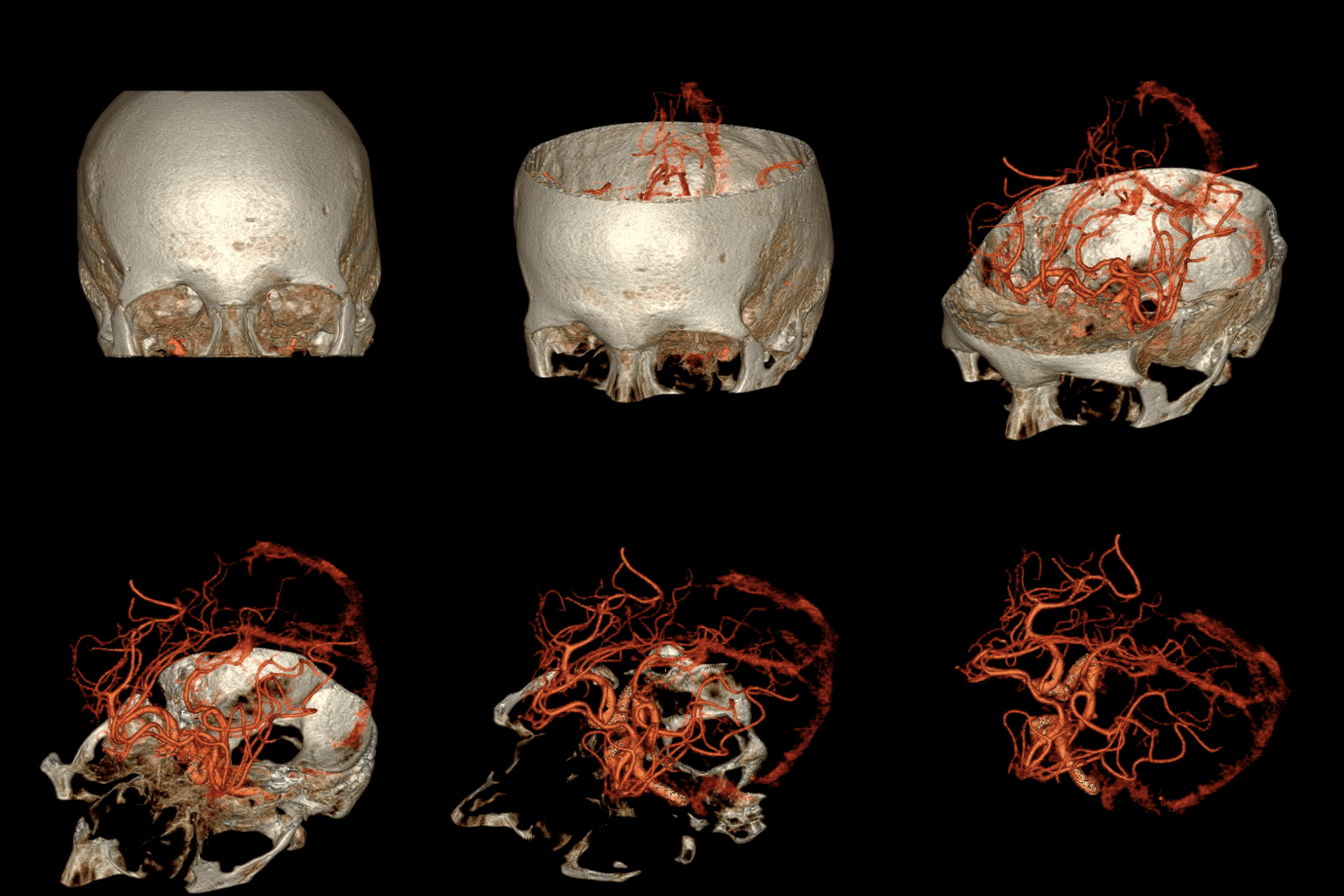
Doctors can use it to observe the pale pink material in the pleura or brain covering the surface of the lungs, and even "enter" a part of the body to check for polyps, tumors and other lesions. This technology helps to provide doctors with more intuitive image information and better preparation for surgery.
Construct a 3D human anatomical model
Currently, 3D imaging has become a frontier in medical imaging technology. Booker's Center of Excellence has pioneered the construction of 3D human anatomical models using images acquired by GE scanning equipment. Doctors can use this model to perform targeted observations on a particular organ, such as coronary stenosis, and to print a convenient organ model using a 3D printer.

Avot and Le Berre have helped VR technology become an important tool in the field of medical diagnostics through the “Open Innovation and Design Thinking Processâ€. Their products have also been recognized by experts.
It is reported that this technology will soon be able to land, and a French customer of GE will try this VR system this year, and then it is expected to be put into use in the US and Asia. The first VR prototype they produced will help doctors gain a better understanding from an anatomical perspective and help doctors better determine the type of pathology. Avot hopes that this technology will not only help doctors diagnose, but also hope that doctors will use the VR model to perform step exercises before surgery, and evaluate the surgical results after surgery to reduce the risk of surgery.
Blowing Mouth,Disposable Blowing Mouth,Filter Spirometer Mouthpiece,Disposable Spirometer Mouthpiece
Hengshui Qifei Paper Products Co. LTD , https://www.hengshuiqifei.com
